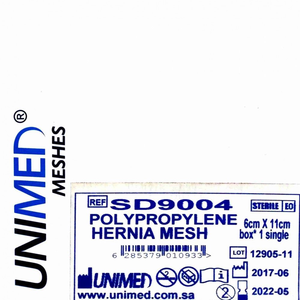
The surgical mesh (in English: Surgical mesh) is a thin loosely woven fabric used either as a permanent or temporary support for organs and other tissues during surgery. The surgical mesh is made of inorganic materials and also biological materials and is used in a variety of surgical procedures. Although hernia repair surgery is the most common application, it can also be used in reconstructive works, such as pelvic organ prolapse. Permanent meshes remain in the body, while temporary meshes dissolve over time. For example, in 2012, testing of the TIGR® Matrix mesh showed that it completely dissolves after three years in a scientific experiment on sheep. Some types of meshes combine being both permanent and temporary, such as Vipro, which contains both absorbable fibrin, made from polyglactin acid, and "Prolene," a non-absorbable polypropylene. Data on the mechanical and biological behavior of meshes in vivo may not always be consistent within the human body, as they are tested in other living organisms. Most published reports have conducted experiments on mice, which raises suspicion of potential differences when implanted in the human body. Also, most published research reports are currently being removed from the medical device market due to post-surgical complications. Additionally, the lack of regulatory protocols approved by the Food and Drug Administration and global standard surgical procedures leads to a variety of different testing methods from researcher to researcher. Some meshes may yield different results in experimentation.
Medical uses
The primary function of the surgical mesh is to support sagging organs either temporarily or permanently. Its most common use is in intra-abdominal hernia surgery, which is required when an organ protrudes through abdominal muscles. The surgical mesh can also be used to reconstruct the pelvis or vaginal wall in women, and it is implanted as an addition to guide the growth of damaged tissue. To be ideal, these implants must be strong enough to withstand mechanical pressures and movements of the body area — whatever it may be — that the mesh becomes part of.
Hernia surgery
Hernia surgery is one of the most common current applications of surgical mesh. Hernias occur when organs or fatty tissues protrude through openings or weak areas in muscles, usually in the abdominal wall. The surgical mesh is implanted to reinforce tissue repair and reduce recurrence rates. Surgery can be performed using laparoscopy (internally) or by open surgery with the use of various available materials for the patching process. Polypropylene is the most used type of mesh, although it may cause discomfort for the patient after implantation. Another less-used type in hernia surgery is polyethylene terephthalate, which faces some complications regarding its tendency to shrink easily after a few years of implantation, nullifying the effects of the surgery. Tetrafluoroethylene is also used, but it is manufactured in the form of metallic chips and faces difficulty in integrating into surrounding tissues, thus lacking stability.
Pelvic surgery
Similar to hernia surgery, manufactured meshes can also be used to treat organ prolapse in the pelvic area. Pelvic organ prolapse occurs in 50% of women over fifty with a history of vaginal delivery once or more during their lifetime. Mesh implantation surgery can be performed in different areas of the pelvis, such as the bladder prolapse, rectal prolapse, vaginal vault, or uterus. The most commonly used material, as in hernia surgery, is polypropylene, which is considered biocompatible in this area. It elicits a mild inflammatory response but has a tendency to adhere to viscera. The vaginal wall consists of three layers: the thick mucous membrane, the muscular layer, and the outer serosa. When prolapse occurs, the smooth fibers of the muscular layer weaken. It has been observed that prolapse in women increases pelvic stiffness, especially after menopause. The surgical mesh used in pelvic reconstruction must resist this stiffness, but if the elasticity coefficient is too high, it will not support the organs adequately. Conversely, if the mesh is too rigid, tissue erosion will occur, and inflammatory responses can lead to post-surgical complications. Additionally, the mesh must have enough strength to withstand basic movements and tissue behavior under physiological conditions, especially during tissue regeneration through the mesh itself. This area is subjected to various loads related to abdominal contents, pressure from abdominal muscles/diaphragm, reproductive organs, and breathing movements. For a woman of reproductive age, the pelvis must withstand a load of 20 Newtons when lying down, 25-35 Newtons when standing, and 90-130 Newtons during coughing. Any implant in the pelvic area must be strong enough to withstand these loads.
Biocompatibility
Implanting the mesh will naturally generate an anti-inflammatory response to the foreign mesh, but the extent of its biological compatibility ranges from ease of integration to the severity of the foreign body reaction. The minimal response involves fibrosis formation around the artificial mesh (similar to scar tissue formation); this response is associated with the best form of biocompatibility. The physical response involves a severe inflammatory reaction, including the formation of giant cells and granulomas, meaning the tissues "accept" the presence of the mesh well. Finally, the chemical response allows for a severe inflammatory reaction during the integration attempt between the mesh and tissues, including increased fibroblast activity. Ultimately, the goal of producing a surgical mesh is to create a mesh with minimal reaction in the living body to maximize patient comfort, avoid infection, and ensure clean integration into the body for tissue repair.
Many factors contribute to the biocompatibility of the mesh. The mesh porosity is the ratio of the pores to the total area and plays a role in the development of bacterial infection or smooth tissue regeneration depending on pore size. Pores smaller than 10 micrometers are susceptible to infection because bacteria can enter and multiply, while macrophages and neutrophils are too large to enter the pores and cannot help eliminate bacteria. Pores larger than 75 micrometers allow fibroblasts, blood vessels, and collagen fibers to pass through as part of tissue regeneration. Although there is no consensus on the optimal pore size, it can be inferred that larger pores are better for tissue development and integration within the living body. It is known that the current problem with a variety of meshes used in all types of surgeries is that they are not sufficiently biocompatible. Polypropylene has proven effective as a mesh for repairing sagging organs but may cause significant discomfort due to its high elasticity coefficient. This makes the implanted mesh rigid and produces a more pronounced inflammatory response, hindering integration with the surrounding tissues. As previously mentioned, tetrafluoroethylene dissolves easily in the body, and tissues face a difficult time integrating with tetrafluoroethylene. For these reasons, researchers have begun exploring different types of surgical meshes that may be more suitable for the biological environment and provide better comfort during support of sagging organs.
Polyvinylidene fluoride (PVDF) non-fibrous mesh
One specific type of mesh under study is polyvinylidene fluoride (PVDF), or the non-fibrous mesh, which has been found to be more resistant to hydrolytic degradation and non-integration, unlike tetrafluoroethylene, it does not increase in stiffness with age, and unlike polypropylene. It has been tested in both hernia and pelvic/vaginal wall surgeries and is produced by layering fibers one over the other, whereas polypropylene is formed through a tissue-like process. This gives the non-fibrous mesh a heavy-weight structure but low porosity, and it also adds more rigidity and high pressure tolerance compared to polypropylene. This is supported by the (HSP 70) baseline — an indicator of cellular stress and protection against damage, which is beneficial for the artificial mesh and tissue formation — which has been observed and noted to be more available in PVDF mesh. Laboratory observations on the non-fibrous mesh showed evidence of cell migration and spreading on the mesh body. Successful cell growth in elongated spindle shapes with clear boundaries has been observed. The major advantage of using non-fibrous mesh is its ability to carry much larger stem cells than traditional polypropylene mesh, which can improve cell-based treatment for organ prolapse and regeneration. Another important feature of PVDF mesh is the formation of new blood vessels after 12 weeks, which is essential for wound healing. Faster formation of new blood vessels leads to quicker tissue repair and regeneration, reducing the likelihood of mesh exposure or rejection. Further improvements are also needed for PVDF mesh before it can be used in human surgeries. Although its elasticity coefficient is higher than polypropylene, the stretch under equal pressure is much smaller, which could cause complications such as tissue atrophy and loss of mechanical strength. Currently, the non-fibrous mesh also induces a greater foreign body reaction and inflammatory response, which are disadvantages for the biological reactivity purpose of the mesh. For these reasons, PVDF mesh remains under study and experimentation for implantation in the body.
Reducing inflammatory response with mesenchymal stem cells
The inflammatory responses to mesh implantation stimulate tissue formation around the mesh fibers and proliferation of fibroblasts, granulocytes, and macrophages, all of which aid in mesh integration. Failure to manage inflammatory responses can lead to foreign body reactions and complete encapsulation of the implanted mesh, which negates any functional purpose it was meant to serve. It is known that mesenchymal stem cells reduce inflammatory responses, and when combined with a surgical mesh, they can potentially make it uncontrollable and difficult to manipulate. Mesenchymal stem cells can be used with surgical meshes as a ready-made product and enhance macrophage polarization in both in vivo and in vitro environments. This can promote an anti-inflammatory response and regulate the inflammatory reaction associated with mesh implantation.